Imagine waking up and seeing a dark, heavy curtain slowly closing over your field of vision. For many, this isn't a nightmare-it's the first sign of a retinal detachment. This is a medical emergency where the retina, the light-sensitive layer at the back of your eye, peels away from its supporting tissue. Because the retina depends on that tissue for blood and nutrients, once it detaches, the photoreceptors begin to die. If you don't get it fixed fast, you could face permanent blindness in that eye.
The good news is that modern medicine is incredibly good at fixing this, provided you act quickly. According to the National Eye Institute, the goal is to reattach the retina before irreversible damage occurs. In fact, some data shows that surgery performed within 24 hours of the first symptom has a 90% success rate in restoring the anatomy of the eye. The clock is your biggest enemy here; every hour you wait can potentially decrease your visual recovery by about 5%.
| Symptom | What it feels like | Urgency Level |
|---|---|---|
| Floaters | Sudden swarm of dark spots or "cobwebs" | High (Warning) |
| Flashes (Photopsias) | Lightning streaks or sparks in peripheral vision | High (Warning) |
| The "Curtain" Effect | A dark shadow creeping across your vision | Critical (Emergency) |
| Blurry Vision | Sudden distortion or loss of focus | Critical (Emergency) |
Spotting the Red Flags
You shouldn't wait for a total blackout to seek help. There are specific warning signs that usually appear before the full detachment happens. The most common is a sudden explosion of floaters. We all have a few floaters as we age, but if you suddenly see dozens of new dark spots or squiggly lines, your retina might be tearing.
Then there are photopsias-those annoying flashes of light that look like camera flashes or lightning bolts, especially when you move your eyes. These happen because the vitreous gel inside your eye is pulling on the retina. The most dangerous sign, however, is the "curtain." If a dark shadow begins to block your peripheral vision and moves toward the center, your retina is actively peeling away. This requires an immediate trip to the ER or an ophthalmologist.
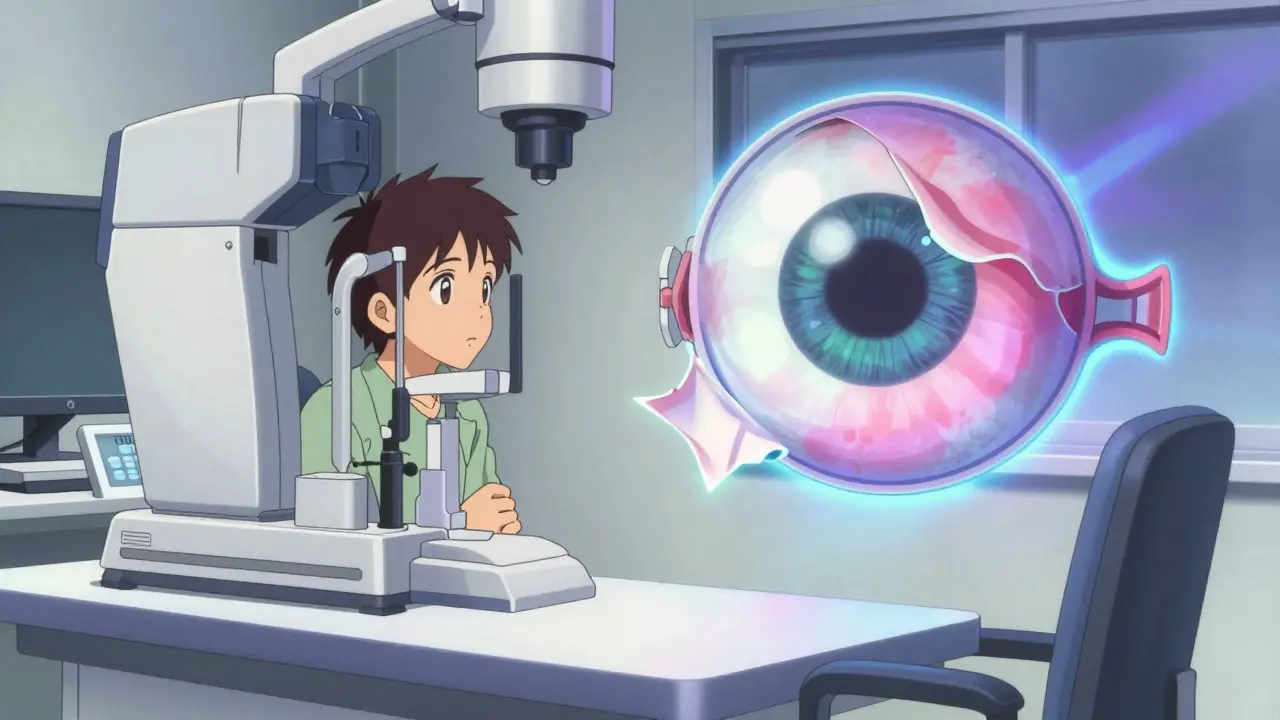
How Doctors Diagnose the Tear
When you get to the clinic, the doctor won't just look at your eye with a flashlight. They need a deeper look. The gold standard is a dilated fundus examination. They use drops to widen your pupil so they can see the entire back of the eye using an indirect ophthalmoscope. This allows them to spot the exact location of the tear.
If your eye is too cloudy for a direct view, they use B-scan ultrasonography. This uses sound waves to create a map of the eye's interior, allowing the surgeon to see if the retina is detached even if they can't see through the pupil. They might also use Optical Coherence Tomography (OCT), which provides a high-resolution cross-section of the retinal layers to see if the macula-the part responsible for your sharp, central vision-is still attached.
Surgical Options: Which One is Right for You?
Not all detachments are treated the same way. The choice of surgery depends on where the tear is and how complex the detachment has become. There are three main paths: pneumatic retinopexy, scleral buckling, and vitrectomy.
Pneumatic Retinopexy is the least invasive. The surgeon injects a gas bubble into the eye that pushes the retina back against the wall. It’s great for single, superior tears (tears at the top of the eye), but it doesn't work for tears at the bottom. It has a decent success rate, but about 30% of patients end up needing a second, more permanent surgery.
Scleral Buckling is like putting a rubber band around the eye. The surgeon places a silicone band (the buckle) around the outside of the eye to push the wall inward until it meets the detached retina. This is often the preferred choice for younger patients or those with lattice degeneration (thinning of the retinal tissue). While it's very effective, it can sometimes cause a slight shift in your prescription, making you slightly more nearsighted.
Vitrectomy is the heavy hitter and the most common procedure today. The surgeon removes the vitreous humor (the gel filling the eye) and replaces it with a gas or oil bubble to hold the retina in place. It's the best option for complex cases or "giant retinal tears." The success rate is very high (90-95%), but there's a catch: it almost always accelerates the formation of cataracts, meaning you'll likely need cataract surgery within a couple of years.
| Method | Success Rate | Best For... | Main Downside |
|---|---|---|---|
| Pneumatic Retinopexy | 70-80% | Single, top-of-eye tears | High re-operation rate |
| Scleral Buckling | 85-90% | Young patients / Lattice degeneration | Potential for myopia/double vision |
| Vitrectomy | 90-95% | Complex detachments / Macula-off | Speeds up cataract development |
Life After Surgery: The Recovery Struggle
The surgery itself is often the easy part; the recovery is where it gets tough. If you have a gas bubble in your eye from a vitrectomy or pneumatic retinopexy, you may be required to maintain a face-down position for several days. Why? Because the bubble needs to float up and push the retina against the back of the eye. If you sit up or sleep on your back, the bubble moves away from the tear, and the surgery could fail.
Patients often report that spending 10 to 20 hours a day looking at the floor is the most grueling part of the process. You might need help at home just to eat or use the bathroom. You'll also need to be careful with travel-you absolutely cannot fly in an airplane or go to high altitudes with a gas bubble in your eye, as the pressure change could cause the bubble to expand and dangerously increase your eye pressure.
Who is at Higher Risk?
While this can happen to anyone, some people are more prone to it. If you are highly nearsighted (myopes with prescriptions greater than -5.00D), your eyeball is often longer, which stretches the retina and makes it thinner and easier to tear. People who have had cataract surgery also have a slightly higher risk because the process can change the dynamics of the vitreous gel.
Then there's lattice degeneration. This is a condition where some areas of the retina are thinner than others. If you've been told you have "lattice" during an eye exam, don't panic, but do stay vigilant. Some doctors suggest treating these thin areas with a laser (prophylactic treatment) before they ever tear, while others prefer to monitor them. Either way, knowing you're at risk means you can spot those first few floaters and get to a doctor before the "curtain" falls.
Can retinal detachment be treated without surgery?
Generally, no. While some very small retinal tears can be "welded" in place using laser therapy or cryopexy (freezing) to prevent a full detachment, once the retina has actually detached from the wall, surgical intervention is required to physically push it back and seal the leak.
Will I get my vision back after surgery?
The primary goal of surgery is to save the eye and prevent total blindness. If the macula (the center of your vision) was still attached when you had surgery, the chances of excellent vision recovery are very high. If the macula had already detached, the retina can be reattached, but your central vision may remain blurry or distorted.
How long does the gas bubble stay in the eye?
It depends on the gas used. Some bubbles dissolve in a few days, while others (like SF6 or C3F8) can take weeks. Your surgeon will monitor the bubble's size during follow-up appointments. Once the bubble is gone, you no longer need to maintain the face-down position.
Is retinal detachment painful?
Surprisingly, the detachment itself is usually painless. You won't feel the retina peeling away. This is why it's so dangerous-people often ignore the visual symptoms because there's no pain to warn them. Any eye-related vision loss should be treated as an emergency regardless of pain.
What happens if I ignore the floaters and flashes?
Ignoring these signs can lead to a full detachment. Once the retina detaches, the photoreceptors are cut off from their blood supply. If you wait several days, the chance of regaining high-quality vision drops significantly. For example, waiting beyond 72 hours can slash the odds of regaining 20/40 vision from 75% down to 35%.